
4.1 – Anatomy and physiology of the large bowel
The large bowel starts at the ileocaecal junction and extends down to the anus. It consists of the caecum, colon, rectum and anus or anal canal.
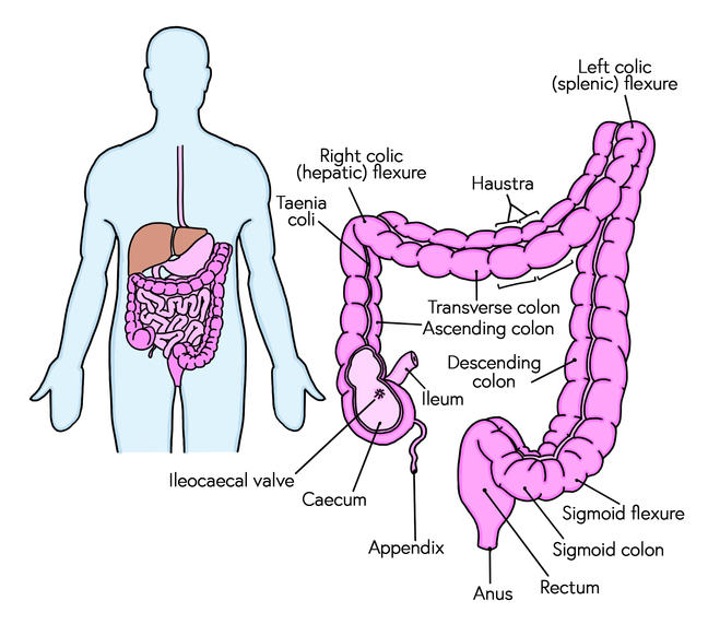 Figure 4.1: Anatomy of the large bowel.
The caecum is the first part of the large bowel, it is joined to the ileum by a sphincter muscle, the ileocaecal valve. The ileocaecal valve is normally constricted therefore preventing contents of the small bowel entering the large bowel and preventing contents of the large bowel going back up into the small bowel.
After a gastrocolic reflex, peristalsis in the ileum is increased, forcing the contents through the valve into the caecum. (We will be discussing the gastrocolic reflex and peristalsis later on in module).
The colon is about 1.5m in length and is divided into four sections:
- the ascending colon 20cm long
- the transverse colon 46cm long
- the descending colon 31cm long
- the ‘S’ shaped sigmoid colon 46cm long.
The hepatic flexure is where the colon turns below the liver to from the transverse colon and the splenic flexure (as it is next to the spleen) is the sharp bend between the transverse colon and the descending colon.
The rectum starts at the end of the sigmoid colon and is 12-15cm in length and has a larger diameter than the rest of the colon which enables it to stretch to accommodate the faeces.
The anus is the last part of the colon, it is the external opening of the rectum